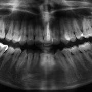
Oral surgery refers to all surgical procedures that must be performed within the oral cavity. Except for rare exceptions, oral surgery is always performed under local anaesthesia.
Oral surgery may be employed for the following reasons:
- Tooth extraction: impacted or non-impacted dental elements
- Odontogenic infections
- Endodontic surgery: apicectomy
- Cysts of the jaw
- Minor pre-prosthesis surgery
- Frenulum
To simplify, we have excluded implantology which is explained further in a separate section.
Tooth Extraction
Teeth that cannot otherwise be recovered due to caries, root fractures, periodontitis or malposition must be removed to avoid causing acute or chronic infections. The teeth that most frequently require removal are the wisdom teeth.
Epidemiological studies show that dysodontiasis of the wisdom teeth is seen in about 20-30% of the population in developed countries and is more prevalent among women. Dysodontiasis (malposition) of the wisdom teeth has a phylogenetic basis: the evolution of an upright posture and the expansion of the brain have led to a reduction in the mandible and maxilla space, also a diet of mainly cooked foods that do not require a third molar. For these reasons, wisdom teeth may sometimes not develop (anodontia) or be positioned incorrectly.
Depending on its location and location in relationship to other significant anatomical structures (inferior alveolar nerve), in addition to orthopantomography (OPT), a CT (Computed Tomography) scan may be performed to choose the least invasive surgical approach.
Possible complications from not extracting wisdom teeth in case of dysodontiasis:
- Pericoronitis: infection of the soft tissues around the crown of the tooth which leads to oedema, localised pain, facial oedema and trismus extending along the muscle mass
- Periodontitis located in the adjacent teeth: sometimes the inflammation affects the second molars, compromising their periodontal health
- Caries: affecting both the wisdom tooth and the roots of the second molar
- Root resorption
- Orthodontic and prosthetic issues
In order to avoid complications, the best age for extraction is between 15 and 20 years old. At this age, the bone is still relatively elastic and the roots are not fully formed with the periodontal ligament sheathe.
The surgery is performed with antibiotic coverage and anti-inflammatories; these are also to be given to the patient following the surgery with directions on how to avoid discomfort in the operated area.
Odontogenic infections. These generally include periapical abscesses that occur with swelling and pain and are associated with mobility of the affected tooth. If untreated, they can develop into a phlegmonous infiltration or the infection can spread through the intraoral soft tissues and muscles.
Surgery includes an incision in the fibro-cellular capsule and draining the abscess, followed by targeted antibiotic therapy.
Apicectomy. When there is a periapical infection (granuloma or abscess) of a tooth that has already been treated with endodontic therapy and it is not possible to extract it because of the presence of endodontic posts or prosthetic crowns, we must resort to endodontic surgery.
The intervention consists of surgical removal of the apex of the tooth and infection, then “retrograde” cleaning of the canal. The canal is then closed with a hydrophilic, mineral trioxide aggregate-based cement.
Cysts of the jaws. These are chronic lesions characterised as a cavity filled with liquid coated with an epithelial wall. Cysts tend to increase in size to the detriment of the anatomical structures surrounding them. Non-odontogenic and pseudocysts are classified as odontogenic cysts (inflammatory and non-inflammatory).
The treatment of choice consists of enucleation of the cyst or complete removal of the cyst in a single operation.
The cyst is then preserved in formalin for histological examination.
Pre-prosthetic surgery. This includes all the interventions necessary to restore the proper anatomy of the soft and hard tissues in order to provide adequate support for the prosthesis.
Frenulum. The frenula are thin bands of soft tissue with a fibro-muscular structure. The three main frenula are the upper and lower labial frenulum, and the lingual frenulum.
Hypertrophic frenulum can lead to functional limitations and diastema of the teeth that can only be treated through surgical removal of the frenulum; the intervention is called a frenulectomy and consists of making an incision and removing the frenulum and, where necessary, removal of the muscle attachments.


