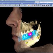
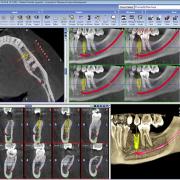
The dental practice is equipped with state of the art, low x-ray emission digital radiology systems; images are scanned through sensors which allow for 3D display.
The following are available:
- Endoral XR (single tooth)
- Panoramic XR (entire mouth including mandible and maxilla)
- Cone beam 3D XR (one or more sections of the oral cavity)
The 3D cone beam system solves all problems related to previous CAT systems (substantial emission of ionizing radiations, slow image scans, feeling of claustrophobia for the patient); 3D cone beam x-rays allows the patient to stand upright with a sensor circling around him for just a few seconds: image scans are immediately available and allow to develop a 3D model of any sector in the oral cavity. The software also allows for a simulation of the operation to be performed, thus minimizing any potential complication.
All systems are periodically controlled by radiologists who certify their proper functioning.


